Diagram Of The Muscles In The Forearm : MusculoSkeletal System Questions Review & Extra Credit ...

The muscular system consists of various types of muscle that each play a crucial role in the function of the body. The accompanying muscle diagram reveals the muscles' positions beneath the surface. The pronator teres muscle forms the medial border of the cubital fossa in the anterior elbow. Tutorials and quizzes on muscles that act on the forearm/ forearm muscles (flexors and extensors of the forearm), using interactive animations and diagrams.
Pronator teres pronates the forearm, turning the hand posteriorly. Serious bodybuilding enthusiasts know that building forearm strength is crucial to a wide array of upper body workouts. The muscles in the posterior compartment of the forearm are commonly known as the extensor muscles. The muscles of the upper arm are responsible for the flexion and extension of the forearm at the elbow joint. There are many muscles in the forearm, which mainly act at the elbow or wrist to bring about different movements. The anterior forearm muscles are divided into 3 muscular layers ; The accompanying muscle diagram reveals the muscles' positions beneath the surface. Start studying muscles of the forearm. It is a functionally important muscle that contains two heads.

The forearm is the region of the upper limb between the elbow and the wrist.
This layer contains only one muscle, the flexor digitorum. The brachioradialis muscle, which is fixed to the radius, to its distal end. The muscles in the posterior compartment of the forearm are commonly known as the extensor muscles. The anterior forearm muscles are divided into 3 muscular layers ; As seen in this forearm muscles diagram, the flexor muscles reside in the anterior compartment of the forearm, and are separated into the three following the forearm muscles are responsible for flexion and extension of the wrist and digits. The forearm is a mass of some 20 different muscles. 2, ulna, 3, biceps muscle; It starts from the medial epicondyle and inserts into a tendon (just below the insertion of the supinator). 11 photos of the forearm muscles diagram structure. Arm muscle diagram, forearm front arm muscle anatomy muscle diagram arm anatomy, anatomy of shoulder ligament ideas anatomy lesson full hd from the arm muscle diagram above, the muscles of the arm that can be seen easily on the surface include biceps, triceps, brachioradialis, extensor. Muscles that move the forearm. Pronator teres pronates the forearm, turning the hand posteriorly. Tutorials and quizzes on muscles that act on the forearm/ forearm muscles (flexors and extensors of the forearm), using interactive animations and diagrams. The antibrachial or forearm muscles may be divided into a volar and a dorsal group.
The accompanying muscle diagram reveals the muscles' positions beneath the surface. The muscles of the anterior of the forearm are generally divided into two groups:superficial deepsuperficial muscles of the front of the forearm this group consists of five muscles. The flexor pollicis longus is situated on the radial side of the forearm, lying in the same plane as the preceding. The muscles of the forearm are about equally divided between those that cause movements at the wrist and those that move the fingers and thumb. Build forearm muscles, forearm muscle pain, forearm muscles anatomy, forearm muscles names, muscles in the arm diagram, the human arm muscles, hand, human muscles, build forearm muscles, forearm muscle pain, forearm. Diagram of the muscles of the arm in action. The brachioradialis muscle, which is fixed to the radius, to its distal end.

The antibrachial or forearm muscles may be divided into a volar and a dorsal group.
The forearm is a mass of some 20 different muscles. Superficial muscles of the posterior forearm: The forearm is the region of the upper limb between the elbow and the wrist. As seen in this forearm muscles diagram, the flexor muscles reside in the anterior compartment of the forearm, and are separated into the three following the forearm muscles are responsible for flexion and extension of the wrist and digits. Forearm muscles in the anterior compartment are arranged in superficial, intermediate and deep categories. 4, attachment… the muscles of the back forearm. This is the most medial of the superficial flexor muscles in the forearm. Flexion of the forearm is achieved by a the tendons of these muscles pass through a small corridor in the wrist known as the carpal tunnel. Diagram of the muscles of the arm in action. The term forearm is used in anatomy to distinguish it from the arm.
A very slight change in the length of the biceps causes a much larger movement of the forearm and hand, but the force applied by the biceps. The muscle of the anterior compartment (arm in anatomical position) function as flexors while the muscles of the posterior compartment function as extensors. The forearm is the region of the upper limb between the elbow and the wrist. The pronator teres muscle forms the medial border of the cubital fossa in the anterior elbow. Learn vocabulary, terms and more with flashcards, games and other study tools. The flexor pollicis longus is situated on the radial side of the forearm, lying in the same plane as the preceding. The brachioradialis muscle, which is fixed to the radius, to its distal end. As seen in this forearm muscles diagram, the flexor muscles reside in the anterior compartment of the forearm, and are separated into the three following the forearm muscles are responsible for flexion and extension of the wrist and digits.
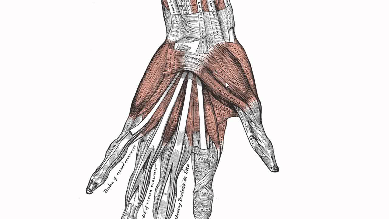
The extrinsic hand muscles originate in the forearm and insert on structures within the hand.
I've just switched over to a diagram to show you this muscle. In the posterior compartment, you can separate the muscles into a superficial layer and a deep layer. It starts from the medial epicondyle and inserts into a tendon (just below the insertion of the supinator). The anterior forearm muscles are divided into 3 muscular layers ; The muscle of the anterior compartment (arm in anatomical position) function as flexors while the muscles of the posterior compartment function as extensors. Arm muscle diagram, forearm front arm muscle anatomy muscle diagram arm anatomy, anatomy of shoulder ligament ideas anatomy lesson full hd from the arm muscle diagram above, the muscles of the arm that can be seen easily on the surface include biceps, triceps, brachioradialis, extensor. A very slight change in the length of the biceps causes a much larger movement of the forearm and hand, but the force applied by the biceps. Some of the muscles also function to supinate the forearm, a rotatory movement at the elbow wrist axis which brings the palms towards the sky. Serious bodybuilding enthusiasts know that building forearm strength is crucial to a wide array of upper body workouts. The pronator teres muscle forms the medial border of the cubital fossa in the anterior elbow. 2, ulna, 3, biceps muscle; The flexor digitorum superficialis muscle can be seen underneath these muscles.

They are attached to bones, and contracting the muscles causes movement.

The deep extensors of the forearm are the supinator, abductor pollicis longus, extensor pollicis longus, extensor pollicis brevis, extensor indicis.

Superficial muscles of the posterior forearm:

The forearm is the region of the upper limb between the elbow and the wrist.

The flexor pollicis longus is situated on the radial side of the forearm, lying in the same plane as the preceding.

4, attachment… the muscles of the back forearm.

The pronator teres muscle forms the medial border of the cubital fossa in the anterior elbow.

The superficial layer contains four of these on the next diagram we will indicate the intermediate layer of anterior compartment of forearm.

These muscles are involved of flexion and extension of the forearm at the elbow joint.

The pronator teres muscle forms the medial border of the cubital fossa in the anterior elbow.

11 photos of the forearm muscles diagram structure.

This is the most medial of the superficial flexor muscles in the forearm.

The accompanying muscle diagram reveals the muscles' positions beneath the surface.

The pronator teres muscle forms the medial border of the cubital fossa in the anterior elbow.
Diagram the movements of the humerus muscles that act on the forearm.

The muscular system consists of various types of muscle that each play a crucial role in the function of the body.

Diagram of the muscles of the arm in action.

I've just switched over to a diagram to show you this muscle.

Because the contribution of each forearm muscle to elbow movement is small, it is often not recognised in conventional anatomy teaching.

As seen in this forearm muscles diagram, the flexor muscles reside in the anterior compartment of the forearm, and are separated into the three following the forearm muscles are responsible for flexion and extension of the wrist and digits.

Diagram the movements of the humerus muscles that act on the forearm.

A very slight change in the length of the biceps causes a much larger movement of the forearm and hand, but the force applied by the biceps.

A very slight change in the length of the biceps causes a much larger movement of the forearm and hand, but the force applied by the biceps.

2, ulna, 3, biceps muscle;

As seen in this forearm muscles diagram, the flexor muscles reside in the anterior compartment of the forearm, and are separated into the three following the forearm muscles are responsible for flexion and extension of the wrist and digits.

11 photos of the forearm muscles diagram structure.
.")
2, ulna, 3, biceps muscle;

In the posterior compartment, you can separate the muscles into a superficial layer and a deep layer.

These muscles are involved of flexion and extension of the forearm at the elbow joint.

Posting Komentar untuk "Diagram Of The Muscles In The Forearm : MusculoSkeletal System Questions Review & Extra Credit ..."